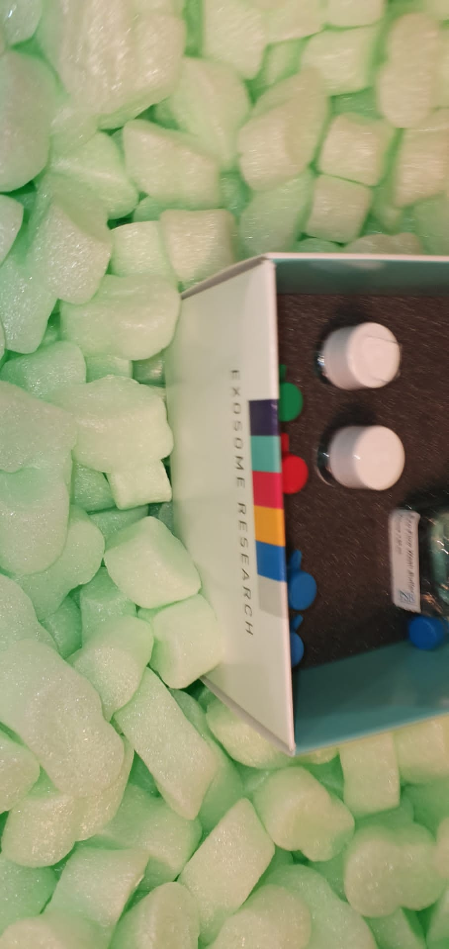
Although cryopreserved cell specimens are used throughout biomedical research, the sample thaw process is high-intensive and prone to an error. Here we describe a small laboratory device that flows a non-frozen cell bottle with a conical tube containing hot cell culture supports. The entire complex is loaded directly into a centrifuge; Less than 5 minutes, cells are thawed and diluted from toxic cryopreservation medium. The recovery and viability of the cells are slightly reduced compared to the common (traditional) method.
However, the antigen-specific T cell function is not affected. Since no technician time is required (beyond the discharge of the vials), our device makes it possible to treat as many samples as the centrifuge can contain (up to 96, in some models). In addition, since the samples are not manually thawed in a water bath, the problems associated with the technician-to-technician differences in the manipulation of the samples are minimized, such as the potential for contamination. It is important to note that the elimination of substantial workforce involving subjective decisions standardize this process and can reduce the variability of the results of cryopreserved specimens.
Currently, the centrifugal apparatus is mainly extremity end sample treatment equipment. The absence of real-time active control has imposed a limitation inherent in such delicate sample processing measures requiring immediate and accurate intervention has never been possible. We note here an engine-assisted chip-in-a-tube platform in which a microfluid chip placed inside a common centrifuge cartridge can be pivoted through the wireless control to handle The centrifugal force vector in a 3-dimensional (3D) in the manner. As a demonstration experience, we used our MACT prototype to perform the operation of two common biomedical procedures, namely the separation of human blood plasma and plasmid DNA extraction E. coli. This simple, but highly efficient and versatile approach can serve as a single generic platform for a wide range of common laboratory experiences and BioAssay applications.
The impact of the characteristics of the centrifuge and centrifugation protocols on cells, growth factors and fibrin architecture of a grass and fibrin membrane rich in leukocytes and ceilings (L-PRF).
The F-PRF (Fibrin rich in leukocytes and platelets) is one of four families of platelet concentrates for surgical use and is widely used in oral and maxillofacial regeneration therapies. The first objective of this article was to evaluate the mechanical vibrations appearing during centrifugation in four commercially available table centrifuge models used to produce L-PRF and the impact of the characteristics of the centrifuge on the cell and fibrin Architecture of an L-PRF clot and membrane. The second objective of this article was to assess how to change some L-PRF protocol parameters may influence its biological signature, regardless of the characteristics of the centrifuge. In the first part, four commercially available centrifuges were used to produce L-PRF, following the original production method L-PRF (glass covered glass tubes, 400 g of force, 12 minutes).
The systems tested were the centrifuge of origin L-PRF (intra-spin, intra-lock, the only EC and FDA erased system for the preparation of L-PRF) and three other laboratory centrifuges (not ceded CE / FDA cleared for L-PRF): A-PRF 12 (PRF Advanced, Process), LW-UPD8 (LW Scientific) and Salvin 1310 (Salvin Dental). Each centrifuge was opened for inspection, two accelerometers were installed (a radial, a vertical) and the data were collected with a spectrum analyzer in two configurations (load at full load or half charge). All clots and membranes were collected in a sterile surgical box (Xpression Kit, intra-lock). The exact macroscopy (weight, sizes) and microscopic (electronic electron microscopy and SEM scanning microscopy) of the L-PRF produced with these four different machines were evaluated.
In the second part, the venous blood was taken from two groups, respectively, 9 ml of plastic glass tubes (intra-lock lock) and 10 ml A-PRF glass (method). The tubes were immediately centrifuged at 2700 rpm (about 400 g) for 12 minutes to produce L-PRF or 1500 rpm for 14 minutes to produce A-FRF. All centrifugations were carried out using the L-PRF (intra-spin) centrifuge, as recommended by both manufacturers. Half of the membranes were placed individually in the culture media and transferred to a new experimental seven-hour tube (up to 7 days). Transformation growth factor β-1 (TGFβ-1), Growth factor derived from AB platelet (PDGF-AB), vascular endothelial growth factor (VEGF) and bone morphogenetic protein 2 (BMP-2) have quantified using ELISA kits at these seven experimental hours.
The remaining membranes were used to evaluate the initial amount of L-PRF and A-FRF membrane growth factors through forced extraction. Very significant differences in the vibration level at each speed of rotation have been observed between the four centrifuges tested. The original L-PRF (intra-spin) centrifuge was by far the most stable machine of all configurations and has always remained below the resonance threshold, unlike the other three machines tested. At the classic speed of L-PRF production, the level of unwanted vibration on the original centrifuge was between 4.5 and 6 times lower than with other centrifuges. Intra-spin showed the lowest temperature of the tubes. A-FRF and Salvin were both associated with a significant increase in temperature in the tube.
Intra-spin produced the heaviest clot and amount of exudation among the four techniques. A-FRF and LW have produced a lot of lighter clots and membranes, shorter and narrower than the other two centrifuges. The analysis of light microscopy has shown relatively similar characteristics for all L-PRF (cellular concentration in the first half). However, SEM has illustrated considerable differences between samples.

