Extreme irritation results in secondary immune harm after traumatic mind damage (TBI). The intestinal mucosa is a key element of immune tolerance because of gut-brain axis regulation, however the healing impact isn’t optimum. Intestinal dysfunction impairs the institution of immune tolerance in sufferers with TBI. Subsequently, we orally administered mind protein (BP) mixed with probiotics to induce immune tolerance and explored the mechanism by which the homeostasis of the microbiota contributes to the enhancement of healing results by BPs. Herein, we demonstrated that sufferers with TBI and surgical mind damage (SBI) fashions of rats had apparent dysbiosis. Notably, the intestinal barrier, proinflammatory cytokines, and activation of microglia demonstrated that extreme inflammatory harm was higher managed within the mixed group (oral administration of BP mixed with probiotics) than within the BP group (oral administration of BP). Basically, tandem mass tag (TMT)-based quantitative proteomics evaluation revealed that BP and probiotics preferentially have an effect on Strive-related pathways.
A sequence of experiments additional confirmed that Indoleamine 2,three dioxygenase (IDO)/Kynurenine (Kyn)/Aryl hydrocarbon receptor (AhR) expression was excessive within the BP group, whereas Tryptophan hydroxylase 1(TpH1)/5-hydroxytryptamine (5-HT) solely modified within the mixed group. This examine means that probiotics can improve the efficacy of oral BP-induced immune tolerance by means of the Strive pathway.
C reactive protein to albumin ratio (CAR) as predictor of anastomotic leakage in colorectal surgical procedure
Background: Anastomotic leakage (AL) is likely one of the most extreme issues in colorectal surgical procedure. At the moment, no predictive biomarkers of AL can be found. The purpose of this examine was to analyze the position of C reactive protein (CRP) to albumin ratio (CAR) as a predictor of AL in sufferers present process elective surgical procedure for colorectal most cancers.
Supplies and strategies: Knowledge on 1183 consecutive sufferers surgically handled for histologically confirmed colorectal most cancers within the surgical items concerned within the examine have been collected. Knowledge included intercourse, age, BMI, ASA rating, Charlson comorbidity index, localization, histology and stage of the illness, in addition to blood exams together with albumin and CRP at the 4th postoperative day. Variations in CAR between sufferers who developed AL and people who didn’t have been analyzed, and the power of CAR to foretell AL was investigated with ROC evaluation.
Outcomes: CAR was considerably greater in sufferers with AL as compared to these with out, on the 4th postoperative day. In ROC evaluation CAR confirmed a great capability in detecting AL (AUC 0.825, 95%CI: 0,786-0,859), larger than these of CRP and albumin alone. CAR additionally confirmed a excessive capability in detecting postoperative deaths (AUC 0.750, 95% CI 0,956-0,987). These findings have been confirmed in multivariate evaluation together with probably the most related threat elements for AL.
Conclusion: Our examine evidenced that CAR, a cheap and extensively accessible laboratory biomarker, adequately predicts AL and demise in sufferers who underwent elective surgical procedure for colorectal most cancers.
The position of TRIM proteins in PRR signaling pathways and immune-related ailments
Sample recognition receptors (PRRs) are a sort of recognition molecules primarily expressed on innate immune cells. PRRs acknowledge a number of sorts of pathogen-associated molecular patterns (PAMPs), inducing the manufacturing of interleukin (IL), tumor necrosis issue (TNF), interferon (IFN) and different associated cytokines to worsen immune-related ailments. PPR signaling pathways play an essential position in each innate and adaptive immune system, and they’re straightforward to be activated or regulated.
Tripartite motif (TRIM) proteins are a bunch of extremely conserved proteins in construction. Most of TRIM proteins comprise RING area, which is believed to play a task in ubiquitination. TRIM proteins are concerned in viral immunity, inflammatory response, autophagy, and tumor progress. On this evaluate, we concentrate on the regulation of TRIM proteins on PRR signaling pathways and their roles in immune-related ailments.

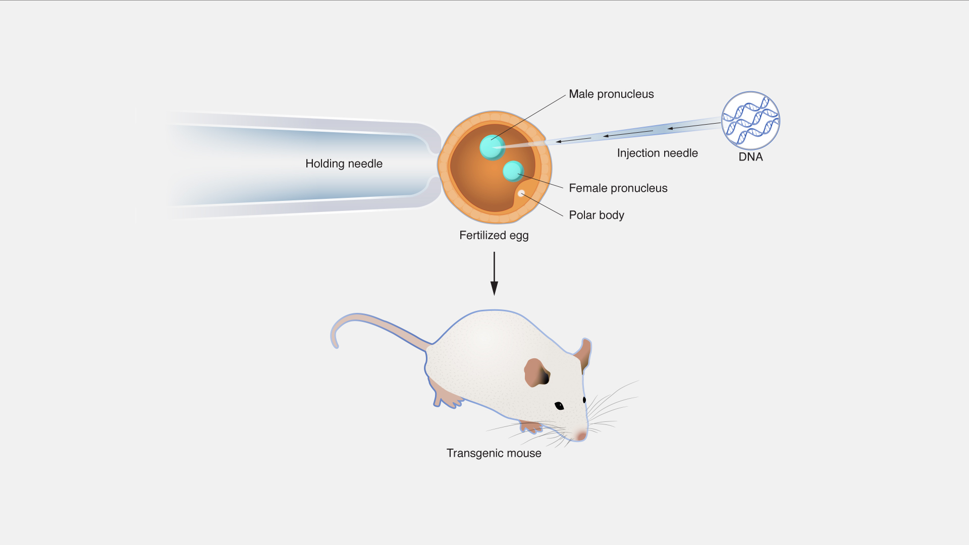
transgenicnews
A fluorescent molecular imaging probe with selectivity for soluble tau aggregated protein
Soluble types of aggregated tau misfolded protein, usually termed oligomers, are thought of to be probably the most poisonous species of the totally different meeting states which are the pathological parts of neurodegenerative issues. Subsequently, a important biomedical want exists for imaging probes that may determine and quantify them. We now have designed and synthesized a novel fluorescent probe, pTP-TFE for which binding and selectivity profiles in the direction of aggregated tau and Aβ proteins have been assessed. Our outcomes have proven pTP-TFE to be selective for early types of soluble tau aggregates, with excessive affinity of dissociation constants (Okay d) = 66 nM, and tenfold selectivity over mature tau fibrils.
Moreover, we discovered that pTP-TFE is selective for tau over Aβ aggregates and had good cell permeability. This selectivity of pTP-TFE in the direction of early types of aggregated tau protein ex vivo was additionally supported with research on human mind tissue containing tau and Aβ pathology. To one of the best of our information, that is the primary fluorescent molecule to be reported to have this type of selectivity profile, which means that pTP-TFE is a singular probe candidate for imaging-based detection of early levels of Alzheimer’s illness and different tauopathies.
Reconstituted cryopreserved platelets synthesize proteins throughout short-term storage and packaging an outlined subset into microvesicles
Background: Cryopreservation of platelets (PLTs) might enable extension of their shelf-life to years, in comparison with days for liquid saved platelets. Resulting from their larger hemostatic impact, reconstituted cryopreserved platelets (cryo-PLTs) would be capable to help bleeding emergencies. Since protein synthesis has been linked to PLT capabilities, reminiscent of clot formation and immune responses, the translational capability of reconstituted cryo-PLTs was assessed upon thawing and short-term storage.
Strategies/supplies: Platelets have been frozen at -80°C with 5-6% DMSO. Upon thawing, they have been reconstituted in plasma after which aliquoted (12 ml) into mini-bags and assessed over 24 h of storage at RT. One sequence served as management; the second and third sequence have been spiked with both 300 μM puromycin (Pm) or 227 nM biotin-labeled Pm. Samples have been examined for in vitro high quality and PLT microvesicle enumeration by movement cytometry. Protein synthesis in cryo-PLTs was assessed utilizing a modified technique based mostly on puromycin-associated nascent chain proteomics.
Outcomes: In vitro parameters of reconstituted and subsequently saved platelets have been in step with beforehand printed outcomes. Mass-spectrometry analyses recognized that 22 proteins have been synthesized in PLTs and 13 of these have been noticed in platelet microvesicles (PMVs).
Conclusion: Cryo-PLTs can synthesize proteins upon reconstitution and storage. Discovery of a subset of those proteins within the PMV suggests a task in vesicle encapsulation, presumably in a selective method. This commentary supplies novel insights into the capability for protein synthesis in cryo-PLTs and the potential regulation of protein packaging into PMV.
![]() Goat anti Mouse IgG1 (Alexa Fluor 488) |
|||
43R-1649 | Fitzgerald | 500 ug | EUR 570 |
Description: Goat anti Mouse IgG1 secondary antibody (Alexa Fluor 488) |
![]() Anti-Hu CD16 Alexa Fluor® 488 |
|||
A4-646-T100 | ExBio | 100 tests | EUR 269 |
![]() Nuclear Pore Complex antibody |
|||
10R-8370 | Fitzgerald | 100 ul | EUR 349 |
Description: Mouse monoclonal Nuclear Pore Complex antibody |
![]() Anti-Nuclear pore Complex antibody |
|||
STJ120173 | St John's Laboratory | 100 µl | EUR 469 |
![]() AF488-streptavidin conjugate [Streptavidin, Alexa Fluor™ 488 Conjugate] |
|||
16891 | AAT Bioquest | 1 mg | EUR 176 |
![]() AF488 Phalloidin [equivalent to Alexa Fluor® 488 phalloidin] |
|||
23153 | AAT Bioquest | 300 Tests | EUR 306 |
![]() Anti-Hu CD72 Alexa Fluor® 488 |
|||
A4-310-T100 | ExBio | 100 tests | EUR 269 |
![]() Anti-Bov CD9 Alexa Fluor® 488 |
|||
A4-354-C100 | ExBio | 0.1 mg | EUR 269 |
![]() Endoglin/CD105 Alexa Fluor |
|||
FC15024 | Neuromics | 100 Tests | EUR 448 |
![]() Panspecific Nuclear Pore Complex Marker |
|||
MO22107 | Neuromics | 500 ul | EUR 344 |
![]() Nuclear pore complex protein Nup153 Antibody |
|||
20-abx109340 | Abbexa |
|
|
![]() Goat Anti-Mouse IgG(H+L) Alexa Fluor 488–conjugated |
|||
S0017 | Affbiotech | 200ul | EUR 304 |
![]() Goat Anti-Rabbit IgG(H+L) Alexa Fluor 488–conjugated |
|||
S0018 | Affbiotech | 200ul | EUR 304 |
![]() SAM FCM (Alexa Fluor 647) |
|||
abx098902-100tests | Abbexa | 100 tests | EUR 1233 |
![]() Anti-LAMP3 antibody (Alexa-fluor 546) |
|||
STJ170005 | St John's Laboratory | 100 µg | EUR 393 |
Description: The dendritic cell lysosomal-associated membrane protein (DC-LAMP)/CD208 is a type I integral transmembrane glycoprotein mostly homologous to CD68, of about 45 kDa in mouse and 90 kDa in human (glycosylation), with a bipartite C-terminal structure divided by a serine/proline rich region, a transmembrane domain and a conserved tyrosine-based lysosomal targeting motif in its cytoplasmic tail. Initially cloned as a specific marker of human mature dendritic cells (DCs), DC-LAMP has been subsequently shown to be expressed in alveolar type II pneumocytes. In both cell types, the molecule is found in the limiting membrane of intracellular multi-lamellar bodies, known as MIIC (MHC class II compartments) in human mature DCs and as lung surfactant-containing lamellar bodies in type II pneumocytes. In the latter cell type, DC-LAMP expression is also detected at the cell surface. |
![]() Anti-LAMP3 antibody (Alexa-fluor 647) |
|||
STJ170006 | St John's Laboratory | 100 µg | EUR 393 |
Description: The dendritic cell lysosomal-associated membrane protein (DC-LAMP)/CD208 is a type I integral transmembrane glycoprotein mostly homologous to CD68, of about 45 kDa in mouse and 90 kDa in human (glycosylation), with a bipartite C-terminal structure divided by a serine/proline rich region, a transmembrane domain and a conserved tyrosine-based lysosomal targeting motif in its cytoplasmic tail. Initially cloned as a specific marker of human mature dendritic cells (DCs), DC-LAMP has been subsequently shown to be expressed in alveolar type II pneumocytes. In both cell types, the molecule is found in the limiting membrane of intracellular multi-lamellar bodies, known as MIIC (MHC class II compartments) in human mature DCs and as lung surfactant-containing lamellar bodies in type II pneumocytes. In the latter cell type, DC-LAMP expression is also detected at the cell surface. |
![]() Anti-IL3RA antibody (Alexa-fluor 546) |
|||
STJ170010 | St John's Laboratory | 100 µg | EUR 393 |
Description: IL3 exerts its biologic activity through its interaction with a cell surface receptor that consists of two subunits. The a subunit (CD123) specifically binds IL3, whereas the ß subunit is required for signaling and is common to the GMCSFR and IL5-R. 107D2.08 and 106C2.02 mAbs were obtained after mouse immunization with sorted human tonsillar PDC. Both clones strongly stain PDCs and basophils, weakly stain monocytes, CD34+ derived DCs and CD11c+ DC, while no staining is observed on T, B, NK cells as well as on mono-derived DCs. Staining with 107D2.08 and 106C2.02 mAbs are maintained on sorted PDC cultured in the presence of IL3 and CD40L, but lost when IL3 alone is added to the culture. The recognition of the IL3Ra chain by 107D2.08 and 106C2.02 was confirmed by transfection studies. 107D2.08 appeared to be the most appropriate clone for in situ studies. 107D2.08 allowed the first observation of IL3Ra+ cells in breast tumor microenvironment |
![]() Anti-IL3RA antibody (Alexa-fluor 647) |
|||
STJ170011 | St John's Laboratory | 100 µg | EUR 393 |
Description: IL3 exerts its biologic activity through its interaction with a cell surface receptor that consists of two subunits. The a subunit (CD123) specifically binds IL3, whereas the ß subunit is required for signaling and is common to the GMCSFR and IL5-R. 107D2.08 and 106C2.02 mAbs were obtained after mouse immunization with sorted human tonsillar PDC. Both clones strongly stain PDCs and basophils, weakly stain monocytes, CD34+ derived DCs and CD11c+ DC, while no staining is observed on T, B, NK cells as well as on mono-derived DCs. Staining with 107D2.08 and 106C2.02 mAbs are maintained on sorted PDC cultured in the presence of IL3 and CD40L, but lost when IL3 alone is added to the culture. The recognition of the IL3Ra chain by 107D2.08 and 106C2.02 was confirmed by transfection studies. 107D2.08 appeared to be the most appropriate clone for in situ studies. 107D2.08 allowed the first observation of IL3Ra+ cells in breast tumor microenvironment |
![]() Anti-CD207 antibody (Alexa-fluor 546) |
|||
STJ170015 | St John's Laboratory | 100 µg | EUR 393 |
Description: Langerin/CD207 is a transmembrane C-type lectin receptor (CLR) of epidermal and mucosal Langerhans cells (LCs) that induces Birbeck's granule formation. Langerin features a single carbohydrate recognition domain (CRD) with mannose-type specificity in its extracellular portion. Langerin is unique among the CLRs in that it contains an intracellular domain with a proline-rich motif. Langerin expression has not been reported outside the DC system. (Valladeau J et al, 1999, Eur.J.Immunol., 29:2695-2704; Valladeau J et al, 2000 Immunity, 12 : 71-81; Kashihara M et al, 1986, J.Invest.Derm., 87 :602-607 Ito T et al, 1999, J.Immunol., 163 :1409-1419 ;Saeland S & Valladeau J, CD207 (Langerin) Workshop reports 2002, Leukocyte-Typing VII, White Cell Diff Antigens, D. Mason et al, Eds, Oxford University Press:306-307) |
![]() Anti-CD207 antibody (Alexa-fluor 647) |
|||
STJ170016 | St John's Laboratory | 100 µg | EUR 393 |
Description: Langerin/CD207 is a transmembrane C-type lectin receptor (CLR) of epidermal and mucosal Langerhans cells (LCs) that induces Birbeck's granule formation. Langerin features a single carbohydrate recognition domain (CRD) with mannose-type specificity in its extracellular portion. Langerin is unique among the CLRs in that it contains an intracellular domain with a proline-rich motif. Langerin expression has not been reported outside the DC system. (Valladeau J et al, 1999, Eur.J.Immunol., 29:2695-2704; Valladeau J et al, 2000 Immunity, 12 : 71-81; Kashihara M et al, 1986, J.Invest.Derm., 87 :602-607 Ito T et al, 1999, J.Immunol., 163 :1409-1419 ;Saeland S & Valladeau J, CD207 (Langerin) Workshop reports 2002, Leukocyte-Typing VII, White Cell Diff Antigens, D. Mason et al, Eds, Oxford University Press:306-307) |
![]() Anti-IL7R antibody (Alexa-fluor 546) |
|||
STJ170021 | St John's Laboratory | 100 µg | EUR 393 |
Description: The IL7-R consists of 2 chains, IL-7R known as CD127 and common cytokine receptor chain known as CD132. A 75 to 80kDa human IL-7 receptor has been cloned that belongs to hematopoietic cytokinereceptor super family. R34-34, raised against human leukemic pre-B cells, recognized a molecule expressed on normal B cell precursors but not on mature B cells. This antibody specifically reverted IL-7 mediated growth inhibition of leukemic BCP (normal B cells precursors) and mature T cells. IL-7R expression is dramatically influenced by cytokines and antigens. This IL-7R displays both high and low affinity for its ligand (IL-7). Inhibitory and proliferative effects of IL-7 can be mediated through the same receptor on various lineages. CD4+ memory T cells express high level of IL-7R Subsets that express it generally require it, including progenitors of T and B cells, naïve and memory T cells. (Pandrau-Garcia D et al, 1994, Blood, 83, 3613-9 Mazzucchelli R et al, Nat. Review Immunol., 2007,7, 144-54) |
![]() Anti-IL7R antibody (Alexa-fluor 647) |
|||
STJ170022 | St John's Laboratory | 100 µg | EUR 393 |
Description: The IL7-R consists of 2 chains, IL-7R known as CD127 and common cytokine receptor chain known as CD132. A 75 to 80kDa human IL-7 receptor has been cloned that belongs to hematopoietic cytokinereceptor super family. R34-34, raised against human leukemic pre-B cells, recognized a molecule expressed on normal B cell precursors but not on mature B cells. This antibody specifically reverted IL-7 mediated growth inhibition of leukemic BCP (normal B cells precursors) and mature T cells. IL-7R expression is dramatically influenced by cytokines and antigens. This IL-7R displays both high and low affinity for its ligand (IL-7). Inhibitory and proliferative effects of IL-7 can be mediated through the same receptor on various lineages. CD4+ memory T cells express high level of IL-7R Subsets that express it generally require it, including progenitors of T and B cells, naïve and memory T cells. (Pandrau-Garcia D et al, 1994, Blood, 83, 3613-9 Mazzucchelli R et al, Nat. Review Immunol., 2007,7, 144-54) |
![]() Anti-Hu CD3 zeta (pY153) Alexa Fluor® 488 |
|||
A4-686-C100 | ExBio | 0.1 mg | EUR 269 |
![]() Anti-Hu CD3 zeta (pY72) Alexa Fluor® 488 |
|||
A4-712-C100 | ExBio | 0.1 mg | EUR 269 |
![]() Anti-Hu CD3 zeta (pY142) Alexa Fluor® 488 |
|||
A4-730-C100 | ExBio | 0.1 mg | EUR 269 |